Screening and diagnostic mammography
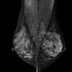
A mammogram involves taking an X-ray image of the breast. This is a leading examination for screening for breast cancer. It allows early detection of cancer, which reduces mortality.
To do this, during the examination, the breast is compressed between two plates, which spreads the breast tissue. An x-ray is then taken. Changes can be observed when there is a potentially neoplastic lesion.

Diagnostic arthrography
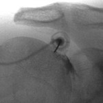
Arthrography is an X-ray examination that involves injecting dye into a joint followed by X-rays.
The patient is lying down, the radiologist applies local anesthesia and then guides his needle with the X-rays until it reaches the correct position. The duration of arthrograms is often less than 15 minutes.

Joint infiltration
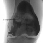
An infiltration consists of injecting a drug. Most often it is cortisone.
Several infiltrations can be done by the attending physician at his office, but when certain points to be infiltrated are too deep or too difficult to access, guidance becomes necessary and radiologists can offer this service.

Magnetic resonance imaging
Magnetic resonance is a medical imaging examination that uses an electromagnetic field and radio frequencies to obtain extremely precise images of the various structures of the human body.
For this, the human body must be in an environment where there is a magnetic field.

General and specialized ultrasound
Ultrasound is a non-invasive and non-irradiating imaging technique that studies the propagation of ultrasound targeted at a particular part of the body.
This examination makes it possible to visualize the soft tissues of the body such as the liver, the spleen, the kidney or the heart. The probe sends ultrasounds of different frequencies to the tissues of the body which will reflect part of these waves.
Bone densitometry
Bone densitometry is a 20-minute test that measures your body's bone mass. Your doctor uses the results to help in his choice of treatment when there is a risk of fracture.

General and dental radiography
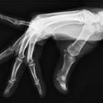
General radiography involves taking images of the internal structures of the body using X-rays.
It is a first-line examination that is quick to perform. Plain x-rays are painless and do not require preparation.
They are performed by professionals in a controlled environment to ensure your safety.
